Brain dysfunction in MCS – Multiple Chemical Sensitivity

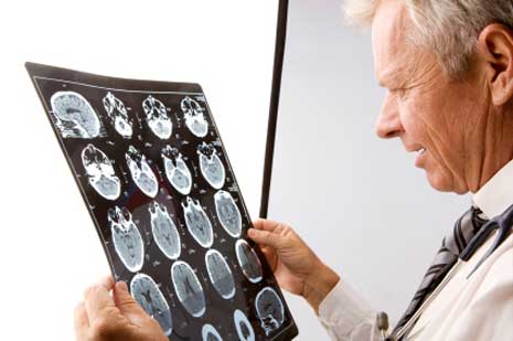
The aim of the following study was to ascertain whether MCS patients present brain single photon emission computed tomography (SPECT) and psychometric scale changes after a chemical challenge.
This procedure was performed with chemical products at non-toxic concentrations in 8 patients diagnosed with MCS and in their healthy controls. In comparison to controls, cases presented basal brain SPECT hypoperfusion in small cortical areas of the right parietal and both temporal and fronto-orbital lobes.
After chemical challenge, cases showed hypoperfusion in the olfactory, right and left hippocampus, right parahippocampus, right amygdala, right thalamus, right and left Rolandic and right temporal cortex regions(p</=0.01). By contrast, controls showed hyperperfusion in the cingulus, right parahippocampus, left thalamus and some cortex regions (p</=0.01). The clustered deactivation pattern in cases was stronger than in controls (p=0.012) and the clustered activation pattern in controls was higher than in cases (p=0.012).
In comparison to controls, cases presented poorer quality of life and neurocognitive function at baseline, and neurocognitive worsening after chemical exposure. Chemical exposure caused neurocognitive impairment, and SPECT brain dysfunction particularly in odor-processing areas, thereby suggesting a neurogenic origin of MCS.
Reference: Orriols R, Costa R, Cuberas G, Jacas C, Castell J, Sunyer J., Brain dysfunction in multiple chemical sensitivity, Servei de Pneumologia, Hospital Universitari Vall d’ Hebron, Barcelona, Catalonia, Spain; CIBER Enfermedades Respiratorias (CIBERES), Spain, J Neurol Sci. 2009 Oct 2.

Leave a Reply
You must be logged in to post a comment.